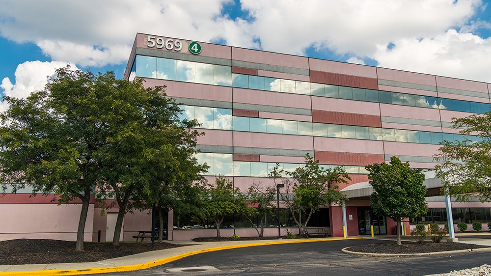
Imaging & Radiology
Mount Carmel offers high-quality outpatient imaging that’s fast, accurate and convenient. With locations at each of our central Ohio hospital campuses, as well as through our growing network of Mount Carmel Imaging Centers and joint-venture locations, we’re close by and easy to find. Having so many American College of Radiology-accredited imaging facilities means we’re also able to offer more appointment options and longer hours – including evenings and Saturdays. And thanks to our state-of-the-art equipment; skilled, licensed radiographers; and board-certified radiologists, you’ll get the best, most precise imaging services available, including:
CT
Computed tomography, or CT, is a special x-ray that uses a scanner and computer to show cross-sectional pictures of structures inside the body, including those in the chest, belly, pelvis, arm or leg, as well as organs like the liver, pancreas, intestines, kidneys, bladder, adrenal glands, lungs, and heart. It also can be used to study blood vessels, bones and the spinal cord.
Interventional Radiology
Interventional Radiology is a rapidly growing area of medicine that uses imaging guidance to perform a wide variety of procedures that would otherwise require more invasive surgery. These minimally invasive, targeted treatments involve no large incisions, which means you’ll generally have less risk, less pain and a shorter recovery time.
MRI
MRI is a noninvasive imaging technique that does not require exposure to radiation. MRI uses a strong magnetic field and radiofrequencies to image the internal organs, soft tissues, brain and spinal cord. As the human body is made up of about 75% water, an MRI uses the water’s hydrogen atoms and computers to produce very detailed images. Learn about what to expect during an MRI.
MRI (Caring Suite)
Mount Carmel East makes MRIs available in the calming environment of our Caring MR Suites. This unique suite allows you to choose a personalized audio/visual experience for your scan — including lighting, music, imagery and video – to make the entire experience more comfortable and less stressful. You can choose a pre-programmed experience like the sights and sounds of nature or create something uniquely personal using photos, videos and music from your own iPhone, iPad or iPod.
Nuclear Medicine
To complete a nuclear medicine scan, you would either swallow or be given a very small dose of a radioactive tracer intravenously. The tracer then accumulates in your tissues (including bones, organs, glands and blood vessels) and makes them more visible so that a technician using a special camera can take images of them.
PET/CT Imaging
A PET scan examines how tissues in the body are using nutrients and to see if cells are normal by injecting a patient with a small amount of a radioactive tracer, similar to nuclear medicine. A CT scan produces images of the body and the organs. A PET/CT scan combines both scans, creating a 3-D image that is often used to detect cancer or changes in the tissues of the heart and brain.
Ultrasound
Ultrasound testing uses high-frequency sound waves to produce images of the organs inside your body. The test is done by moving a device called a transducer over a part of the body. The transducer sends out sound waves, which bounce off the tissues inside your body and bounce back to create the images. It’s commonly used to view organs like the uterus, bladder, kidneys and heart.
X-ray (General Diagnostics)
X-rays are used to view bones and soft tissue inside the body. An x-ray is produced when a small amount of radiation passes through the body and strikes a plate or detector placed on the other side. An x-ray’s ability to penetrate tissue and bone varies depending the mass and composition of the tissue, resulting in a lighter or darker image.


